Although it may currently be impossible to accurately identify individual hairs and their colors, ultrasounds in later stages of pregnancy can still show hair to someone with the proper expertise.
Standard 2D ultrasounds depict hair as thin wisps or a fuzzy halo. On 3D and 4D ultrasounds, hair will appear as wavy contours on your baby’s head.
According to Prenatal Ultrasounds,
“The best method to ‘see’ if the baby has hair is to use the standard, more traditional 2D ultrasound (black and white) which displays ‘fuzz’, a key indication that hair is present.”
While my babies were bald at birth, my niece was born with a head full of thick, black hair that was clearly visible in ultrasounds around the third trimester.
Table of Contents
Hair on Ultrasounds
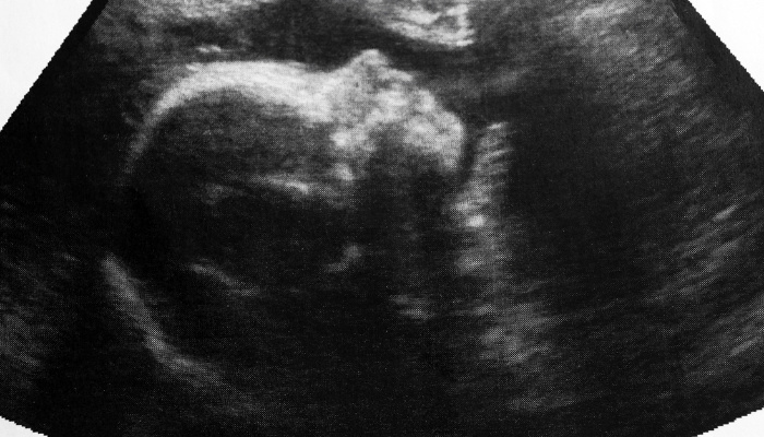
To the untrained eye, an ultrasound is just blobs of black and white, but someone who knows what they’re looking for can see all sorts of things like a baby’s heart, spine, and even hair.
What hair looks like on the screen will depend on what kind of ultrasound you are receiving.
Hair on 2D Ultrasound
Hair is visible on a 2D ultrasound in the later stages of pregnancy.
This hair typically either looks like thin white strands or wisps or a fuzzy halo atop the baby’s head.
What the hair looks like will ultimately depend on the digital clarity of the ultrasound and the amount of hair.
It can be difficult to spot, but a trained sonographer should be able to point out the baby’s hair if any is present.
Experts have greater success seeing hair on a 2D ultrasound rather than a 3D or 4D ultrasound.
Hair on 3D and 4D Ultrasound
3D ultrasounds operate differently than 2D ultrasounds by emitting multiple waves from different angles to produce a 3-dimensional image.
Instead of being able to see individual hairs, you will only see hair on a 3D ultrasound if your baby has a lot of it.
Rather than viewing via color contrast as you would in the 2D ultrasound, hair in a 3D ultrasound looks like an out-of-place contour on the baby’s head.
Don’t worry though, distortion of features and details is typical with 3D ultrasounds.
The main difference between 3D and 4D ultrasounds is that with 4D ultrasounds, you’re able to see your baby move in real time.
The advantage of this is that the baby may be viewed from different angles.
However, looking for something as small as hair on a 4D ultrasound while the baby is moving poses a challenge.
When Hair Becomes Visible on Ultrasound
The time at which hair becomes visible on an ultrasound depends heavily on the amount of hair the baby has.
The baby’s hair follicles are developed around week 15. As their hair begins to grow through these follicles, it becomes increasingly visible.
Hair is often able to be seen at the 20-week anatomy scan.
While you might see hair on an ultrasound, it is not clear enough to predict how much hair your baby will have at birth.
Fetal hair growth is dependent on genetics and maternal hormones during pregnancy.
It is normal for some babies to be born without hair either due to genetics or lack of estrogen.
Hair Color on Ultrasound
You will not be able to see your baby’s hair color on an ultrasound. Even if you could, their hair color will likely change in the first few months of life.
On a 2D ultrasound, hair strands will appear bright white in contrast to the darker background. In 3D ultrasounds, hair is not colored at all.
Factors That Limit Hair Visibility on Ultrasound
There are a few factors that may make it difficult or impossible to see hair on an ultrasound including:
- Amniotic fluid: The amniotic fluid surrounding the fetus can cause distortion in the ultrasound image, making it challenging to visualize small and subtle structures like hair.
- Gestational age: If the pregnancy is still early in gestation, you may not see hair on an ultrasound.
- Fetal Position: Sometimes you are unable to see hair due to the position of the fetus in the womb.
- Hair Thickness: Fetal hair, especially lanugo, is very fine and may not be easily detected by ultrasound.
- Hair Development: Hair development occurs at different rates, and not all fetuses will have significant hair at the time of the ultrasound.
- Type of Imaging Device: Newer, higher-resolution technology can pick up more fine details such as hair while it may be more difficult to spot using older devices.
How To Increase Baby Hair Growth During Pregnancy
Fetal hair growth is driven by estrogen. Eating foods that increase the production of estrogen in the body during pregnancy can positively impact hair growth.
The best foods for this are nuts, seeds, berries, sweet potatoes, avocados, fish, eggs, and spinach.
Fun Fact:
An old wives’ tale says that heartburn during pregnancy indicates that your baby will have a lot of hair.
While there may be a correlation, hair does not cause heartburn. Rather, the hormones that cause hair to grow are what triggers heartburn.
What To Expect To See on Baby Ultrasound
Ultrasounds can pick up several components of your baby’s anatomy and physiology.
Ultrasounds detect both external and internal structures of the body.
For example, it outlines the legs, feet, and toes, but it can also see the bones within them.
In addition, ultrasounds can spot organs, blood vessels, muscles, joints, and tendons.
Ultrasounds are also useful for the early detection of infections, tumors, cysts, and cardiovascular abnormalities.
Issues with fetal anatomy are typically detected at the 20-week anatomy scan if they are present.
When You Typically Get Ultrasounds During Pregnancy
A healthy, uncomplicated pregnancy usually requires 1-2 ultrasounds.
The first ultrasound, held between 7 and 10 weeks, is used primarily for fetal dating and heartbeat detection.
The second ultrasound, between 18 and 22 weeks, is to check the fetal anatomy for abnormalities, infections, and growth.
A complicated or high-risk pregnancy will often require more frequent ultrasounds during the first and third trimesters.
Lanugo vs. True Baby Hair
Lanugo and true baby hair are two different types of hair that infants can have during different stages of development.
Lanugo
Lanugo is a very fine, soft, and downy type of hair that covers the body of a developing fetus.
It usually appears around the 20th week of gestation and begins to shed in preparation for birth.
Lanugo serves a developmental purpose, helping to regulate the temperature of the fetus in the womb.
In most cases, lanugo is shed before or shortly after birth. By the time a full-term baby is born, lanugo is typically no longer present on the body.
True Baby Hair
True baby hair is the more noticeable, coarser, and pigmented hair that develops after the shedding of lanugo.
True baby hair starts to grow after lanugo has shed, and it usually becomes more apparent in the weeks and months following birth.
Baby hair serves some protective and sensory functions, and it continues to develop into the infant’s characteristic hair.
The growth of hair varies among infants. Some may be born with a significant amount, and others may have very little.
The color and texture of true baby hair can also change as the child grows.
Why Some Babies Are Born Without Hair
Hair development in the womb is a complex process, and the timing of hair growth can vary among babies.
The amount and thickness of hair a baby has at birth are influenced by genetics.
If both parents have less hair or thinner hair, it’s more likely that their baby will be born with less hair as well.
Similarly, if both parents have a lot of hair, the baby may be born with a fuller head of hair.
Hair growth patterns can be influenced by factors such as hormones, nutrition, and overall fetal development.
Remember that the amount of hair at birth is not a reliable indicator of future hair growth or characteristics.
Babies’ hair can change significantly in the months following birth, and their final hair texture, color, and thickness may not be apparent until later in childhood.
Charley is a mother of three with a passion for raising good humans. With her children in tow, she studies English and has made a career creating content about motherhood. In her free time, she enjoys traveling within the states to kayak, camp, and hike.